 Search
Determining Normal Fetal Situs (Situs Solitus)
One of the first steps in obtaining cardiac views is to determine how the fetus is oriented within the uterus and to determine the right and left side of the abdominal contents versus the right and left side of the heart and thoracic contents. This is not as easy as it might seem since the fetal left side can be on the maternal right and the opposite can be true.
In summary, situs refers to the right and left orientation of fetal organs. For example situs solitus is the normal left to right axis arrangement in the fetus with the stomach and spleen on the left side of the body, and the liver and gallbladder on the right side.

Above. Situs solitus demonstrating some of the main vascular, cardiac, and pulmonary arrangements.
Visual Summary of Normal Fetal Situs
Below are steps required to determine situs related to cephalic or breech presentation, and whether the spine or back is up (anterior) or down (posterior).
1. Determine the lie of the fetus:
A. Is the fetus head first with the head in front of the ultrasound screen? This could also be termed cephalic or vertex presentation.
B. Is the fetal feet or bottom first with the head behind the screen? This could also be termed footling or breech presentation.
C. Determine whether the spine or back is anterior (back up) or posterior (back down).
2. Obtain a transverse cut of the thorax to demonstrate a 4-chamber view. The left atrium is nearest the spine and the cardiac axis points to the left.
In summary if the fetus is head first (cephalic) and the back is down, on a transverse view of the fetal abdomen the stomach should be on the left.
In summary if the fetus is breech presentation and the back is down, on a transverse view of the fetal abdomen the stomach should be on the right.
Normally, with either presentation, on a 4-chamber view, the left atrium is nearest to the spine.
Detailed Method to Determine Fetal Situs
1. Define within the uterus the presentation of the fetus (generally vertex or breech; less often the presentation is oblique or transverse.).
2. Determine whether the fetal spine is parallel or transverse to the maternal spine. In sagittal view, if the fetal and maternal spine are parallel, the fetus is in longitudinal lie. When the fetal spine is perpendicular to the maternal spine, the fetus is in a transverse lie.
3. Determine the position of the fetal left side. The fetal left side will be as follows:
A. With respect to the maternal abdomen, the fetal left side is anterior and near to the ultrasound transducer.
B. With respect to the posterior uterine wall, the fetal left side is posterior and farthest from the transducer.
C. With respect to the right uterine wall, the fetal left side will be on the maternal right.
D. With respect to the left uterine wall, the fetal left side will be on the maternal left.
4. Obtain a transverse view of the abdomen and define the fetal stomach which is positioned in the left side of the fetal abdomen.
5. Obtain a 4-chamber view of the heart by obtaining a transverse view of the thorax. The left atrium and descending aorta are nearest to the spine and the cardiac axis points to the left.
6. Finally, ascertain if the stomach and heart are in their correct respective locations, i.e., the stomach is on the left side and the cardiac axis points to the left.
7. Place a transverse image of the fetal abdomen and heart side by side and validate that the left side of the fetal abdomen (stomach near to the spine) is concordant with the left side of the fetal heart (left atrium and descending aorta near to the spine). This is done by displaying a side by side comparison of a transverse view through the fetal stomach and a 4-chamber cardiac view.

Above. Potential maternal orientation and transducer relative depth in defining the fetal left side.

Above. Normal ultrasound orientation for situs solitus.
Right Hand Rule of Thumb: Introduction
In their article “Sonographic definition of the fetal situs,” Bronshtein, Gover, and Zimmer [1] describe a “right hand rule of thumb” to define fetal situs during transabdominal scanning, and a “left hand rule of thumb” for transvaginal scanning.

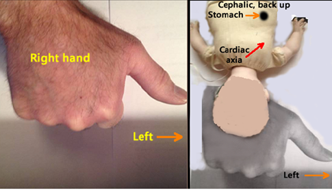
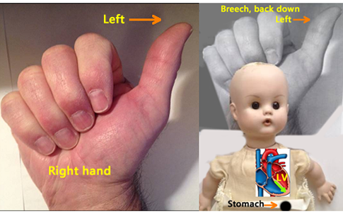
Above. For transabdominal scanning, the right hand is a fist which represents the fetal head. The outstretched thumb always points to the fetal left. The top (dorsal surface) of the hand represents the posterior aspect of the fetus or back up, while the palm side of the fist represents the anterior aspect of the fetus or abdomen, face up with the back down.
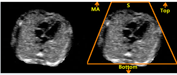
Above. Abdomen. The ultrasound scan plane is from top to bottom with the MA (maternal abdomen) at the top of the screen and ultrasound beams (S) are directed from the transducer as indicated.
The right hand rule of thumb is further illustrated by a the following images:

Above. Cephalic presentation. Right hand rule of thumb demonstrating relative maternal and fetal positions.

Above. Breech presentation. Right hand rule of thumb demonstrating relative maternal and fetal positions.
Right Hand Rule of Thumb: Cephalic, supine, back down

Above. Cephalic presentation, back down. The scan plane is from top to bottom.
Left. The sonographer’s right hand represents the fetus with the thumb pointing to the fetal left. The palm of the hand is anterior, or represents the ventral or face surface of the fetus. The fetus is therefore face up, back down, and the thumb points to the fetal left.
Right. Again, the imaginary fetus is back down with the stomach and cardiac axis pointing to the left. (Ignore color scheme of fetal heart and vessels.)

Above. Ultrasound image. Scan with cephalic presentation (supine) back down. The stomach is on the fetal left and the left atrium is nearest to the DA (descending aorta) and the spine. The axis of the heart is to the left. (Images courtesy of Jill Beithon RT, RDMS, RDCS, RVT).
Right Hand Rule of Thumb: Cephalic, prone, back up
Above. Cephalic presentation, back up. The scan plane is from top to bottom.
Left. The sonographer’s right hand represents the fetus with the thumb pointing to the fetal left. The top of the hand (dorsal surface or prone position) represents back up.
Right. The imaginary fetus is back up with the stomach and cardiac axis pointing to the fetal left.

Above. Ultrasound image. Scan with cephalic presentation (prone) back up. The stomach is on the fetal left and the left atrium is nearest to the DA (descending aorta) and the spine. The axis of the heart is to the left. (Images courtesy of Jill Beithon RT, RDMS, RDCS, RVT).
Right Hand Rule of Thumb: Breech, supine, back down
Above. Breech presentation, back down. The scan plane is from top to bottom.
Left. The sonographer’s right hand represents the fetus with the thumb pointing to the fetal left. The palm of the sonographer’s hand is anterior, or represents the ventral surface of the fetus. The fetus is therefore face up, back down, and the thumb points to the fetal left.
Right. The imaginary fetus is back down with the stomach and cardiac axis pointing to the left. (Ignore color scheme of fetal heart and vessels).

Above. Ultrasound image. Scan with breech (supine) back down. The stomach is on the fetal left and the left atrium is nearest to the DA (descending aorta) and the spine. The axis of the heart is to the fetal left side.
Right Hand Rule of Thumb: Breech, prone, back up

Above. Breech presentation, back up. The scan plane is from top to bottom.
Left. The sonographer’s right hand represents the fetus with the thumb pointing to the fetal left. The top of the hand (dorsal surface or prone position) represents back up.
Right. The imaginary fetus is back up with the stomach and cardiac axis pointing to the fetal left side.

Above. Ultrasound image. Scan with breech presentation (prone) back up. The stomach is on the fetal left and the left atrium is nearest to the DA (descending aorta) and the spine. The axis of the heart is to the fetal left.