 Search
Transvaginal Ultrasound: Images


Above. This image represents a normal transvaginal cervical length with a proper sagittal view, distinct appearance of the distal cervix, and proper placement of the cursors for measurement.

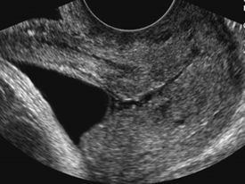
Above. Cervical shortening on transvaginal ultrasound, and visualization of the amnion and chorion.


Above. Spontaneous change in apparent cervical length over a 2 minute observation interval.

Above. The membranes are filling the upper vagina. The approximate internal cervical os dilatation is 2.5 cm, and the approximate external cervical os dilatation is 5.2 cm.

Above. Breaking of the internal cervical os, and demonstration of the amnion and chorion.

Above. U-shaped funnel measuring 20 mm and cervical length measuring 21 mm.

Above. V-shaped funnel with short cervix. The distal cervix is poorly visualized in this image.

Above. An example of a transabdominal ultrasound of the cervical length in the presence of a distended maternal bladder. Compared to the transvaginal approach, this technique is not recommended to obtain accurate cervical length.

Above. Short cervix of 9.8 mm with cerclage sutures intact.

Above. This is a transabdominal cerclage image. The mersilene tape ligature is echogenic and there is no significant cervical shortening. Some ultrasound labs outline the endocervical canal, as illustrated, in the presence of a significantly curved canal, but preference should be given to measurement between cursors, except in extreme cases.