 Search
This educational support is developed for those taking the Limited Obstetric Ultrasound Examinations by Advanced Clinical Providers.
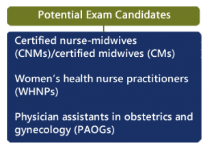
The American Institute for Ultrasound in Medicine (AIUM) has published “AIUM Practice Parameter for the Performance of Limited Obstetric Ultrasound Examinations by Advanced Clinical Providers”.

Obimages.net has compiled a listing of resources from obimages.net for the certifying examination which may help the applicant to achieve competency. An outline from the AIUM document is provided to link the educational chapters to the practice parameters.
We encourage the student to:
- Thoroughly familiarize yourself with the “AIUM Practice Parameter for the Performance of Limited Obstetric Ultrasound Examinations by Advanced Clinical Providers”.
See:
https://www.aium.org/resources/guidelines/LimitedOB_Providers.pdf
To see more information about the examination go to:
https://www.ardms.org/get-certified/midwifery/
The following is a summary and outline of knowledge Areas from “AIUM Practice Parameter for the Performance of Limited Obstetric Ultrasound Examinations by Advanced Clinical Providers”.
Outline of AIUM Practice Parameters
Note Definitions:
As Low as Reasonably Achievable (ALARA) Principle
ALARA principle (as low as reasonably achievable) should be observed when adjusting controls that affect the acoustic output and by considering transducer dwell times. The lowest possible ultrasonic exposure setting should be used to gain the necessary diagnostic information.
A thermal index for soft tissue (TIS) should be used. Recommended Maximum Scanning Times for Displayed Thermal Index (TI) Values
A thermal index for soft tissue (TIS) should be used at earlier than 10 weeks’ gestation, and a thermal index for bone (TIB) should be used at 10 weeks’ gestation or later when bone ossification is evident. A ratio of less than 0.7 is considered appropriate.
Statement on Measurement of Fetal Heart Rate
M-mode imaging or a 2D video clip should be used for documentation of cardiac activity.
Statement on the Safe Use of Doppler Ultrasound During 11–14 Week Scans (or earlier in pregnancy)
In keeping with the ALARA principle, Pulsed Doppler ultrasound is discouraged.
Prudent Use in Pregnancy
Obstetric ultrasound examinations should only be performed when there is a valid medical reason. The use of ultrasound to solely determine fetal sex is inappropriate
Keepsake Fetal Imaging
The promotion, selling, or leasing of ultrasound equipment for making “keepsake fetal videos” is considered by the US Food and Drug Administration to be an unapproved use of a medical device. Use of a diagnostic ultrasound system for these purposes may be in violation of state laws or regulations.
Practice Parameters
The practice parameter is limited to singleton pregnancies. If a multiple gestation is unexpectedly identified, membrane characteristics should be recorded with representative images and the patient should be referred for a standard diagnostic or detailed ultrasound evaluation.
Incidental findings of potential clinical significance should prompt consultation with a physician who at minimum meets the AIUM Official Statement Training Guidelines for Physicians Who Evaluate and Interpret Diagnostic Obstetric Ultrasound Examinations.
Written Request
The written or electronic request for an ultrasound examination should provide sufficient information to allow for the appropriate performance and interpretation of the examination.
The following is a Classification of Fetal Ultrasound Examinations Within the Scope of Practice for Advanced Clinical Providers
I. Limited First Trimester Ultrasound
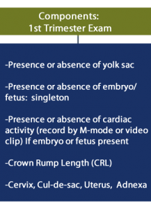
A limited obstetric ultrasound examination in the first trimester includes evaluation of the presence, size, location, and number of gestational sac(s). The gestational sac is examined for the presence of a yolk sac and embryo/fetus. When an embryo/fetus is detected, the crown-rump length (CRL) should be measured and cardiac activity recorded by M-mode imaging or a 2-dimensional (2D) video clip. Pulsed Doppler ultrasound should not be used in the first trimester to “hear” the embryonic heartbeat. The uterus, cervix, adnexa, and cul-de-sac region should be examined.
OBIMAGES.NET FREE CHAPTER DEVOTED TO THIS TOPIC.
https://obimages.net/free-chapter-normal-abnormal-first-trimester-exam/
FIRST TRIMESTER AIUM REQUIREMENTS
- Limited First-Trimester Ultrasound Examination
Indications include but are not limited to:

In addition, adjunct ultrasound guidance for chorionic villus sampling, embryo transfer, and localization and removal of an intrauterine contraceptive device.
- Imaging Parameters
Scanning in the first trimester may be performed either transabdominally or transvaginally. If a transabdominal examination is not definitive, a transvaginal scan is recommended. In some cases, a transabdominal examination may be needed if a transvaginal scan is not definitive.
- The uterus (including the cervix) should be evaluated for the presence of a gestational sac. If a gestational sac is seen, its location should be documented. The gestational sac should be evaluated for the presence or absence of a yolk sac and embryo. If an embryo or fetus is identified, the CRL should be measured.
Comment:
A definitive diagnosis of intrauterine pregnancy can be made when an intrauterine gestational sac containing a yolk sac or embryo/fetus with cardiac activity is visualized.
A small intrauterine fluid collection with an echogenic rim can be seen in the decidualized endometrium. In the absence of ultrasound signs of ectopic pregnancy, such a fluid collection is highly likely (>99.5%) to represent an intrauterine gestational sac. A follow-up ultrasound examination and/or serial determination of maternal serum human chorionic gonadotropin levels are appropriate in pregnancies of undetermined location to avoid inappropriate intervention in a potentially viable early pregnancy.
Although unlikely, an intrauterine fluid collection could represent a “pseudo–gestational sac” associated with an ectopic pregnancy.
If an intrauterine pregnancy is not definitively identified, consultation with a physician provider who at minimum meets the AIUM Official Statement Training Guidelines for Physicians Who Evaluate and Interpret Diagnostic Obstetric Ultrasound Examinations is recommended.
The mean gestational sac diameter is not recommended for estimating a due date. The CRL is a more accurate indicator of gestational age than is the mean gestational sac diameter. The CRL should be measured in a standardized manner.
Table 1. Components of a Limited First-Trimester Obstetric Ultrasound Examination of a Singleton Fetus Presence and location of gestational sac.
ALARA (See above definitions.) TIS at less than 10 weeks’ gestational age; TIB at 10 weeks’ gestation or greater (ratio <0.7).
II. Limited Second or Third-Trimester Examination
A limited obstetric ultrasound examination in the second or third trimester may be performed to answer a specific clinical question: for example, cardiac activity or fetal presentation. A limited obstetric ultrasound examination may also be performed in patients requiring serial examinations in which a subsequent anatomic evaluation may be unnecessary or impractical.
A limited obstetric ultrasound examination does not include an evaluation of fetal anatomy, and in almost all cases, a standard diagnostic or detailed anatomic evaluation of the fetus has been or will be performed during the index pregnancy.
A limited second- or third-trimester ultrasound examination includes an evaluation of fetal number, cardiac activity, presentation, placental location with respect to the internal cervical os, and amniotic fluid volume. If requested, a limited obstetric ultrasound examination may include fetal biometry. Reliable fetal biometric measurements require anatomic familiarity with the midline falx, thalami, cavum septi pellucidi, columns of the fornix, cerebellum, stomach, umbilical vein as it courses through the liver, and femoral diaphysis.
OBIMAGES.NET FREE CHAPTERS DEVOTED TO THIS TOPIC:
https://obimages.net/free-chapter-transducer-placement-fetal-orientation-introduction-heart-exam/
https://obimages.net/free-chapter-normal-fetal-ultrasound-biometry/
https://obimages.net/free-chapter-normal-cns-ultrasound-brain-anatomy/
III. Biophysical Profile
A biophysical profile (BPP) may be performed to assess fetal well-being. The ultrasound component monitors fetal movement, fetal tone, fetal breathing movements, and amniotic fluid volume. This study may be done in conjunction with fetal heart rate monitoring.
A biophysical profile (BPP) may be done to assess fetal well-being. This examination is generally performed in the later second or third trimester in patients at increased risk for antepartum stillbirth:
- Fetal breathing movements—at least 1 episode of rhythmic fetal breathing of 30 seconds or longer within 30 minutes;
- Fetal body movements—at least 3 discrete body or limb movements within 30 minutes;
- Fetal tone—at least 1 episode of extension of a fetal extremity with return to flexion or opening and closing of a hand; and
- Amniotic fluid volume—a single DVP of fluid that does not include the cord or fetal parts (extremities), measuring at least 2 cm in depth and 1 cm in horizontal width.
Each of the 4 areas is given a score of 2 points if the above criteria are met or a score of 0 if the criteria are not met, for a possible total of 8 points. A score of 8 is reassuring. A score of 6 is equivocal (neither reassuring nor nonreassuring). A score of 4 or less is abnormal. Regardless of the BPP score, an inadequate amount of amniotic fluid (single DVP< 2 cm X 1 cm without fetal parts) requires further evaluation. Clinical management depends on gestational age and obstetric circumstances.
While the links to content on obimages.net is thorough, it is not intended to be the only resource for students to prepare themselves for the examination.