 Search
Dextro-Transposition of the Great Arteries (D-TGA): Imaging Considerations and Images
Summary
In dextro-transposition of the great arteries (D-TGA), the four chamber view is normal. However, the five chamber view leads to the diagnosis. If the great vessel arising from the left ventricle branches, the pulmonary artery (PA) is defined and the great vessels are likely transposed with the aorta (AO) arising from the right ventricle instead of the left ventricle.
Dextro-transposition of the great arteries (D-TGA). Patient 1.
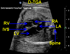

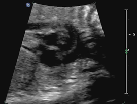
Above. Dextro-transposition of the great arteries (D-TGA). Patient 1. Four chamber view. Note normal four chamber view with the morphologic LV (left ventricle) and RV (right ventricle), and the IVS (ventricular septum) separating the ventricles. The RA (right atrium) and LA (left atrium) are unremarkable. The situs is normal and there is levocardia. Note the normal relationship between the spine and the LA.


Above. D-TGA. Patient 1. Five chamber view. Again, the LV (left ventricle) and RV (right ventricle) appeared normal, but the GA (great artery) arising from the left ventricle branches, likely representing the left branch of the pulmonary artery.


Above. D-TGA. Patient 1. Five chamber view. The great vessel arising from the LV (left ventricle) is clearly the PA (pulmonary artery) dividing respectively into the left and right pulmonary arteries (LPA and RPA). This key view defines relevant anatomy for D-TGA.


Above. D-TGA. Patient 1. Oblique view. The AO (aorta) arises from the RV (right ventricle) and the AV (aortic valve) is noted. A portion of the LV (left ventricle) is seen.


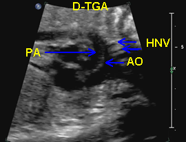
Above. D-TGA. Patient 1. Parasagittal view. The AO (aorta) can be defined by the HNV (head and neck vessels). Note that the AO is the anterior vessel, which is usual in D-TGA. The PA (pulmonary artery) and AO are parallel. A portion of the inflow anatomy is also seen with the RA (right atrium), the RV (right ventricle), and the intervening tricuspid valve. The pulmonary artery appears as a straight course artery and the lateral branching helps to define it. The head and neck vessels arise from the aortic arch.
Dextro-transposition of the great arteries (D-TGA). Patient 2.

Above. Dextro-transposition of the great arteries (D-TGA). Patient 2. Five vessel view. The LV (left ventricle) and RV (right ventricle) are in their normal anatomic position and are morphologically normal. However, the PA (pulmonary artery) arises from the LV and this is defined by the Br. PA (branching of the pulmonary artery). The AO (aorta) arises from the RV.

Above. D-TGA. Patient 2. Same patient showing more detail of the PA (pulmonary artery) arising from the LV (left ventricle). Note the PV (pulmonary valve) and the branching of the PA, which defines this great vessel. The situs is normal and the spine confirms levorotation (leftward), confirming the morphologic LV.


Above. D-TGA. Patient 2. Same patient showing more detail of the AO (aorta) arising from the RV (right ventricle). Note the region of the AV (aortic valve). The situs is normal and the DA (descending aorta) supports LV and RV orientation.
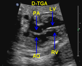

Above. D-TGA. Patient 2. Axial view. Note the parallel arrangement of the great vessels with the PA (pulmonary artery) arising from the LV (left ventricle) and the AO (aorta) arising from the RV (right ventricle). Again, the spine and descending aorta as well as the morphologic characteristics of the ventricles confirm orientation.


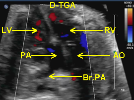
Above. D-TGA. Patient 2. Five vessel view. Color Doppler during systole demonstrating the LV (left ventricle) and the RV (right ventricle) and the great vessels – the PA (pulmonary artery) and the AO (aorta) – arising from the LV and RV respectively. Again, the Br. PA (branching pulmonary artery) helps define this vessel.
Above. D-TGA. Patient 2. Sagittal view of the aorta and the aortic arch. Note the HNV (head and neck vessels) defining the AO (aorta), which is anterior to the PA (pulmonary artery).