 Search
Gastroschisis: Imaging Considerations
The following are ultrasound criteria for the diagnosis of gastroschisis:
• Small bowel present in amniotic fluid.
• Umbilical cord inserts normally (Use Color Doppler).
• No covering membrane.

Above. About 10% of infants with gastroschisis have other malformations. [1] Other large series (334,262) report slightly higher malformation rates of 16.6% [2] Overall, about 50% of the patients with gastroschisis had malformations related to the abdominal wall defect while half of the group had defects not associated with gastroschisis. [1]

Above. Gastroschisis may also be considered as multiple congenital anomalies (MCA). Four hundred and sixty-nine non-isolated cases of gastroschisis were registered in one study. [3] There were 41 chromosomal syndromes, 24 other syndromes, and 404 with multiple congenital anomalies (MCA). [3]
Among those with multiple congenital anomalies, four groups were most frequent: 1) Central nervous system, 4.5%; 2) Cardiovascular system, 2.5%; 3) limb abnormalities, 2.2%; and 4) kidney anomalies, 1.9%. [3]
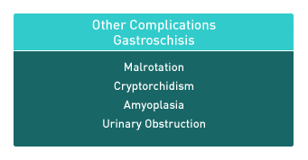
Above. Other malformations specifically related to gastroschisis include: 1) malrotation; 2) cryptorchidism; 3) amyoplasia; 4) urinary tract obstruction; and intestinal atresia or stenosis [4]

Above. Ultrasound findings related to prognosis for gastroschisis include gastric dilatation, other bowel dilatation, polyhydramnios, fetal growth restriction, and other malformations.
In 34 infants with gastroschisis, 21 had no gastric dilatation and 13 had gastric dilatation. Those with gastric dilatation had a higher probability for neonatal death, volvulus, and prolonged hospital care. [5] Both gastric dilatation and large defect are important predictors of outcome. [6]
Abnormal dilatation of fetal bowel of 6 mm to 35 mm was associated with slower oral feeding, decreased primary closure, and increased readmission for bowel obstruction. [7]
Some reports consider the presence of intestinal atresia to be the single most important finding in those with poor outcomes. [8]
Magnetic resonance imaging (MRI) provides a potentially improved contrast without the risk of radiation effect and has been used in anomalies such as congenital diaphragmatic hernia and gastroschisis. [6]
The possibility of 3-D mode in MRI makes the assessment of fetal bowel condition potentially more accurate.