 Search
Placenta Accreta: Images


Above. Transvaginal ultrasound image in patient with previous C-section and placenta. Note small lacunae and and the placental location over the internal cervical os (complete placenta previa).

Above. Transvaginal ultrasound demonstrates placental lacunae in a patient with placenta accreta.
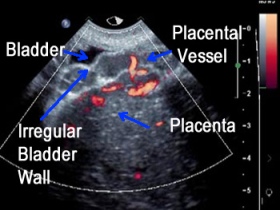
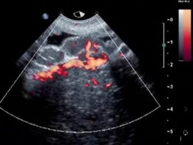
Above. Note precise color Doppler imaging demonstrates increased vascularity. There is increased vascularity and penetration of placental vessels to the myometrium.


Above. Color Doppler imaging demonstrates increased vascularity, and potential penetration of placental vessels to the myometrium.


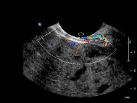
Above. Note relatively smaller lacunae in a patient at risk for placenta accreta.


Above. Note relatively thin myometrium of 1.8 mm in a patient at risk for placenta accreta.

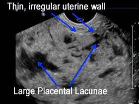
Above. Note the thin uterine wall and the large lacunae suggestive of placenta accreta.